-
NiFe University_of_Regensburg UnivMaryland Cancer Sio2 Lateral_Force_Microscopy block_copolymer SelfAssembly Topography Lanthanum_aluminate VerticalPFM ring shape SThM Mapping BiVO4 Polyaniline Beads GalliumPhosphide ThinFilm strontiu_titanate SiliconeOxide ShenYang AM_KPFM AEAPDES ScanningThermalMicroscopy Tungsten_disulfide Bmp Magnetostrictive Mosfet Wang Optic self-assembled_monolayer plastics molecular_self_assembly align
Report image
If you found this image unacceptable, please let us know. We will review your report and take action if we determine this image is really unacceptable.
2L-MoS₂ (1/3)
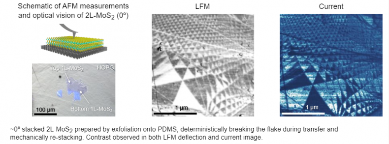
Scanning Conditions
- System : FX40
- Sample bias: 0.25 V
- Scan Mode: C-AFM, LFM
- Scan Rate : 4 Hz
- Scan Size : 2.5μm×2.5μm
- Pixel Size : 512×512
- Cantilever : ContSCPt (k=0.2N/m, f=25kHz)